


Professional career
Urs Eisenmann is the head of the Image-Based Diagnostic and Therapy Support Working Group at the Institute for Medical Informatics at Heidelberg University Hospital. He is also the Quality Management Officer at the Institute for the Development of Software as a Medical Device. Following his studies in Medical Informatics at Heidelberg and Heilbronn universities, he completed his doctorate in computer-assisted neurosurgery at Heidelberg University and worked as a research associate on numerous research projects.
Expertise
The working group is currently focusing on two areas. Prototype tools for computer-assisted surgery are being developed in cooperation with clinics at Heidelberg University Hospital. Within the DZL environment, image-based analysis methods for lung imaging (currently primarily MRI) are being researched. For example, segmentation methods for lung lobes are being developed. Furthermore, the group is researching automation methods for established radiological scoring procedures for various lung diseases (e.g. CF, COPD, PCD and CTEPH). The working group uses image processing techniques, as well as various supervised and unsupervised AI methods.
- Data science and computer-assisted surgery
- Application of image processing and AI methods
- Segmentation + Classification of lung MRI
- Cystic Fibrosis
- Platform Imaging
- DSWG Artificial Intelligence and Digital Tools
- Hagen N, Freudlsperger C, Kühle RP, Bouffleur F, Knaup P, Hoffmann J, Eisenmann U. A User-Friendly Software for Automated Knowledge-Based Virtual Surgical Planning in Mandibular Reconstruction. J Clin Med. 2025 Jun 25;14(13):4508. doi: 10.3390/jcm14134508.
- Ringwald FG, Wucherpfennig L, Hagen N, Mücke J, Kaletta S, Eichinger M, Stahl M, Triphan SMF, Leutz-Schmidt P, Gestewitz S, Graeber SY, Kauczor HU, Alrajab A, Schenk JP, Sommerburg O, Mall MA, Knaup P, Wielpütz MO, Eisenmann U. Automated lung segmentation on chest MRI in children with cystic fibrosis. Front Med (Lausanne). 2024 Nov 12;11:1401473. doi: 10.3389/fmed.2024.1401473.
- Ringwald FG, Martynova A, Mierisch J, Wielpütz M, Eisenmann U. Explainable Artificial Intelligence for Deep-Learning Based Classification of Cystic Fibrosis Lung Changes in MRI. Stud Health Technol Inform. 2024 Jan 25;310:921-925. doi: 10.3233/SHTI231099.
- Kuehle R, Ringwald F, Bouffleur F, Hagen N, Schaufelberger M, Nahm W, Hoffmann J, Freudlsperger C, Engel M, Eisenmann U. The Use of Artificial Intelligence for the Classification of Craniofacial Deformities. J Clin Med. 2023 Nov 14;12(22):7082. doi: 10.3390/jcm12227082.
- Hagen N, Weichel F, Kühle R, Knaup P, Freudlsperger C, Eisenmann U. Automated calculation of ontology-based planning proposals: An application in reconstructive oral and maxillofacial surgery. Int J Med Robot. 2023 Dec;19(6):e2545. doi: 10.1002/rcs.2545.
Prof. Petra Knaup | PI | Petra.Knaup@med.uni-heidelberg.de |
Urs Eisenmann | Group Leader | Urs.Eisenmann@med.uni-heidelberg.de |
Anna Martynova | PhD Student | Anna.Martynova@med.uni-heidelberg.de |
Friedemann Ringwald | PhD Student | Friedemann.Ringwald@med.uni-heidelberg.de |
Niclas Hagen | Postdoc | Niclas.Hagen@med.uni-heidelberg.de |
Lung Research - Projects
1. Deep Learning-Based Classification of Pulmonary Perfusion Defects in Cystic Fibrosis (CF), Chronic Thromboembolic Pulmonary Hypertension (CTEPH), and Chronic Obstructive Pulmonary Disease (COPD) Using Thoracic Magnetic Resonance Imaging (DeePerfusionMRI)
To support the radiological assessment of pulmonary perfusion defects, an AI-based approach is developed for the automated classification of an established scoring system. Due to the lobe-based methodology, preprocessing requires precise lung lobe definition, which is particularly challenging with MRI data. An automated algorithm will be developed for this purpose using image processing and neural networks. In addition to MRI images, clinical data will be integrated to increase the robustness and accuracy of predictions. The focus is on the clinical conditions CF, CTEPH, COPD, and PCD. Patient data will be collected retrospectively at three university hospitals. Explainable AI methods are used to ensure transparency, and evaluation is performed by radiologists.
2. Deep-learning-based classification of structural and functional lung abnormalities in cystic fibrosis based on magnetic resonance imaging (CF-MRI)
In 2012, a lobe-based scoring system was developed at the TLRC to systematically record disease-relevant MRI findings in cystic fibrosis. This system quantifies structural and functional changes in the lungs. To support the assessment, an AI-based approach is being developed to enable automated classification of the scores. The basis is approximately 850 standardized MRI examinations from approximately 200 patients, which were previously assessed using the visual CF-MRI score. Convolutional neural networks are used for training, validation, and testing. The goal is to develop an objective, quantitative measure of morphofunctional changes and a user-friendly decision-making tool for radiologists.
3. Deep Learning-Based Visualization of Perfusion Defects to Support MRI-Based Lung Perfusion Scoring in Cystic Fibrosis (CF-MRXAI)
In 2012, a scoring system was developed at the TLRC to systematically record disease-relevant MRI findings in cystic fibrosis. This system semi-quantitatively captures structural and functional changes. The 4D perfusion sequence with contrast agent, in particular, provides important information on lung perfusion, but is associated with high intra- and inter-reader variability. To date, no satisfactory computer-assisted solutions for quantitative analysis exist. The CF-MRXAI project aims to develop a deep learning-based software tool that supports radiologists with explainable AI methods and enables more objective assessments. In addition to the current sequence, comparable reference cases are visualized. The evaluation is carried out by radiological specialists, with the potential for expansion to include other relevant subscores.
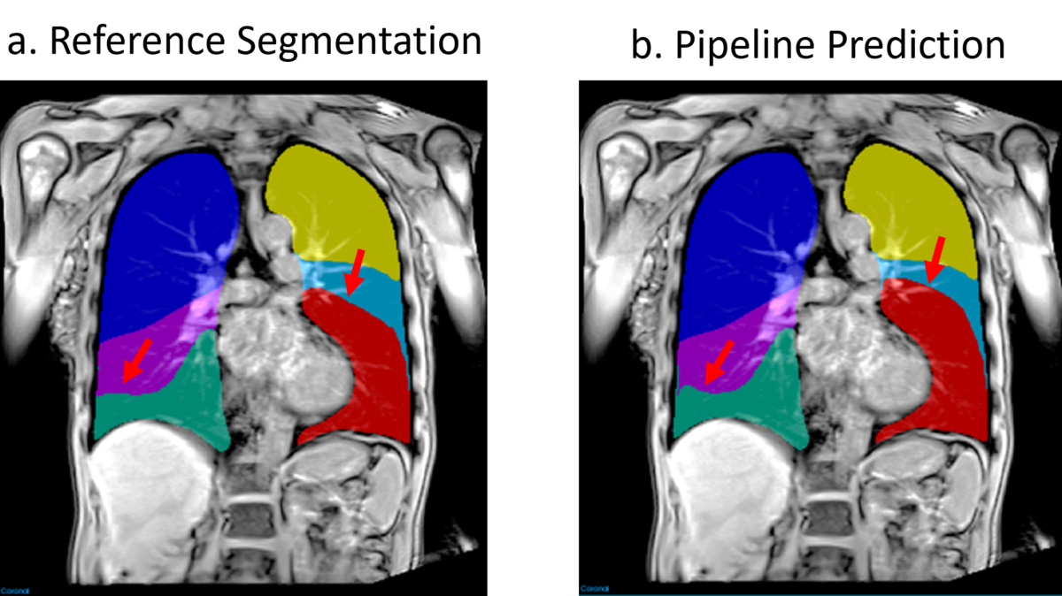

This work presents a deep learning pipeline for automated classification of bronchiectasis/wall thickening and mucus plugging on lung lobes in BLADE MRI data. The approach leverages both axial and coronal image streams, which undergo preprocessing and segmentation to isolate lung halves and lobes. The segmented volumes combined with lobe scores are fed into a dual-stream convolutional neural network with a VGG16 backbone, where features are combined using a late fusion strategy to improve classification performance.